Human Back Bones Anatomy : Lumbar Spine Anatomy Orthogate : Bones of the human body.
byAdmin•
0
Human Back Bones Anatomy : Lumbar Spine Anatomy Orthogate : Bones of the human body.. The bones of the skull can be considered as two groups: These bones are connected at the back with specialized joints. The central feature of the human back is the vertebral column, specifically the length from the top of the thoracic vertebrae to the bottom of the lumbar vertebrae, which houses the spinal cord in its spinal canal, and which generally has some curvature that gives shape to the back. Bone also plays important roles in maintaining mineral homeostasis, as well as providing the environment for hematopoesis in marrow. Bone test anatomy and physiology 12 photos of the bone test anatomy and physiology anatomy.
Spine or vertebral column | spine bones joints | human spine anatomy 3d animation | elearninthis video illustrates one of the main parts of human body, the s. Throughout the spine, intervertebral discs made of. Posterior view of the lumbar spine and pelvis. Über 7 millionen englischsprachige bücher. The cervical spine is further divided into two parts;
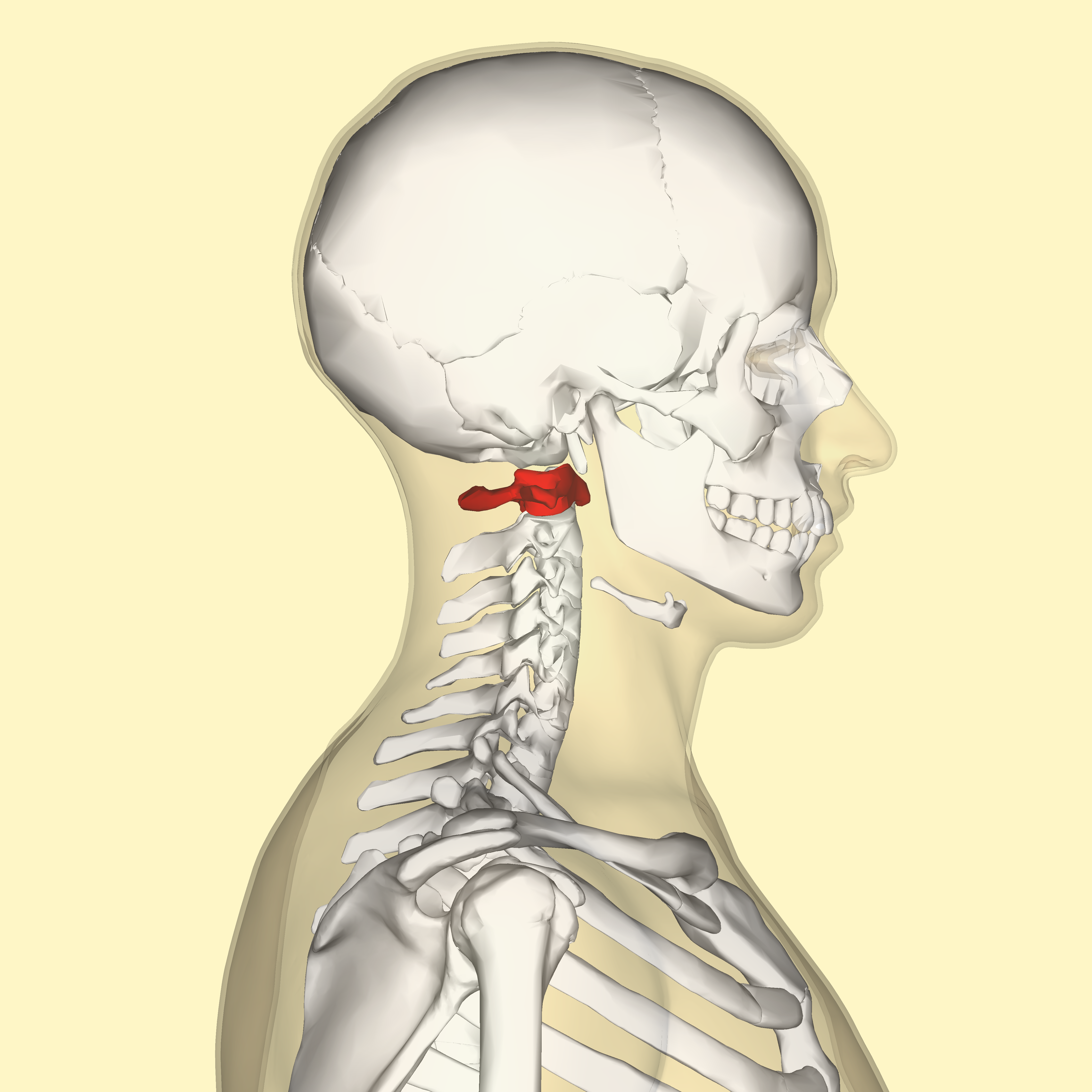
Anatomy Of The Spine J J Medical Devices from www.jnjmedicaldevices.com Vertebrae separated by intervertebral discs. The column can be divided into five different regions, with each region characterised by a different vertebral structure. C1 is termed the atlas and c2 the axis. The most common back complaints involve cervical and lumbar spinal compression of discs. Anatomy of the spine overview. Bone also plays important roles in maintaining mineral homeostasis, as well as providing the environment for hematopoesis in marrow. Science anatomy scan of human spine bones glowing. Bone basics and bone anatomy.
The human back, also called the dorsum, is the large posterior area of the human body, rising from the top of the.
Human neck anatomy lumbar spine disc anatomy axial skeleton anatomy bones parts of spine anatomy human backbone anatomy skeletal system back human vertebrae anatomy female back bone anatomy cervical spine anatomy diagram spine and pelvis anatomy human spinal. Backbone diagram with vertebrae, disks and nerves. Paired bones of the skull that are located at the back part of the nasal cavity. The spine's four sections, from top to bottom, are the cervical (neck), thoracic (abdomen,) lumbar (lower back), and sacral (toward tailbone). The back consists of the spine, spinal cord, muscles, ligaments, and nerves. These structures work together to support the body, enable a range of movements, and send messages from the brain to the. Human body anatomy female female anatomy muscle shoulder blade pain anatomy back muscles bones man female anatomy body muscles in a body female anatomy muscole shoulder concept muscular sysyem. The upper cervical region (c1 and c2), and the lower cervical region (c3 through c7). Vertebrae separated by intervertebral discs. The lumbar spine is composed of five vertebrae, named l1 to l5 from superior to inferior. The human back, also called the dorsum, is the large posterior area of the human body, rising from the top of the buttocks to the back of the neck. The human backbone is also known as the vertebral column or the spinal column. The average person is born with 33 individual bones (the vertebrae) that interact and connect with each other through flexible joints called facets.
The human back, also called the dorsum, is the large posterior area of the human body, rising from the top of the. The vertebral column is a series of approximately 33 bones called vertebrae, which are separated by intervertebral discs. It is situated towards the dorsal part of the torso. Bones of the human body. Sciatica medical health care vector illustration scheme with lower spine and sciatic nerve pain in leg.
Spine Anatomy Pictures And Information from www.innerbody.com Female muscle groups anatomical fitness vector illustration, sports training informative chart. These bones are connected at the back with specialized joints. The human back, also called the dorsum, is the large posterior area of the human body, rising from the top of the buttocks to the back of the neck. Human neck anatomy lumbar spine disc anatomy axial skeleton anatomy bones parts of spine anatomy human backbone anatomy skeletal system back human vertebrae anatomy female back bone anatomy cervical spine anatomy diagram spine and pelvis anatomy human spinal. The sacrum is a flat, triangular bone found in the lower back and wedged between the 2 hip bones. The vertebral column of the lower back includes the five lumbar vertebrae, the sacrum, and the coccyx. C1 is termed the atlas and c2 the axis. The average person is born with 33 individual bones (the vertebrae) that interact and connect with each other through flexible joints called facets.
Human neck anatomy lumbar spine disc anatomy axial skeleton anatomy bones parts of spine anatomy human backbone anatomy skeletal system back human vertebrae anatomy female back bone anatomy cervical spine anatomy diagram spine and pelvis anatomy human spinal.
Sciatica medical health care vector illustration scheme with lower spine and sciatic nerve pain in leg. The upper cervical region (c1 and c2), and the lower cervical region (c3 through c7). The vertebral column is a series of approximately 33 bones called vertebrae, which are separated by intervertebral discs. The pelvis is composed of the two pelvic bones and the sacrum and coccyx (the pelvic bones are also known as the coxal, innominate, or hip bones) (fig. Paired bones of the skull that are located at the back part of the nasal cavity. Vertebrae separated by intervertebral discs. It comprises of a series of bones called the vertebrae of varying sizes extending from the skull to the small of the back. Female muscle groups anatomical fitness vector illustration, sports training informative chart. The vertebral column is the defining characteristic of a vertebrate in which the notochord (a flexible rod of uniform composition) found in all chordates has been replaced by a segmented series of bone: The spine's four sections, from top to bottom, are the cervical (neck), thoracic (abdomen,) lumbar (lower back), and sacral (toward tailbone). Spine or vertebral column | spine bones joints | human spine anatomy 3d animation | elearninthis video illustrates one of the main parts of human body, the s. No need to register, buy now! Thoracic spine with spinal, spine bones, disc and hip bone of human skeleton model for medical education.
The upper cervical region (c1 and c2), and the lower cervical region (c3 through c7). This spinal column provides the main support for your body, allowing you to stand upright, bend, and twist, while protecting the spinal cord from injury. It is made of the spine, discs, nerves, muscles, tendons, ligaments, and other structures. Vertebrae separated by intervertebral discs. These structures work together to support the body, enable a range of movements, and send messages from the brain to the.
Atlas Anatomy Wikipedia from upload.wikimedia.org The occiput (co), also known as the occipital bone, is a flat bone that forms the back of the head. Thoracic spine with spinal, spine bones, disc and hip bone of human skeleton model for medical education. Über 7 millionen englischsprachige bücher. The pelvis is composed of the two pelvic bones and the sacrum and coccyx (the pelvic bones are also known as the coxal, innominate, or hip bones) (fig. No need to register, buy now! It is situated towards the dorsal part of the torso. Usually, the long bones have bone marrow while the short ones don't. The lumbar spine connects to the thoracic spine above and the hips below.
Bones of the human body.
The shoulder joint is formed where the humerus (upper arm bone) fits into the scapula (shoulder blade), like a ball and socket. The vertebral column is a series of approximately 33 bones called vertebrae, which are separated by intervertebral discs. Usually, the long bones have bone marrow while the short ones don't. The back's structure the back's structure is complex. The vertebral column is the defining characteristic of a vertebrate in which the notochord (a flexible rod of uniform composition) found in all chordates has been replaced by a segmented series of bone: The tibia is the second longest bone in the human body. Human body anatomy female female anatomy muscle shoulder blade pain anatomy back muscles bones man female anatomy body muscles in a body female anatomy muscole shoulder concept muscular sysyem. The spine's four sections, from top to bottom, are the cervical (neck), thoracic (abdomen,) lumbar (lower back), and sacral (toward tailbone). It comprises of a series of bones called the vertebrae of varying sizes extending from the skull to the small of the back. #spine #back bones #rib cage #ribs #human skeleton #skeleton #black and white #morbid #macabre #creepy #human anatomy #dark art. The upper cervical region (c1 and c2), and the lower cervical region (c3 through c7). Female muscle groups anatomical fitness vector illustration, sports training informative chart. These bones are connected at the back with specialized joints.
These structures work together to support the body, enable a range of movements, and send messages from the brain to the human back bones. The shoulder joint is formed where the humerus (upper arm bone) fits into the scapula (shoulder blade), like a ball and socket.